Ultrastructure changes in BMP-2-treated SHED cultured on glycerol preserved human amniotic membrane
Abstract
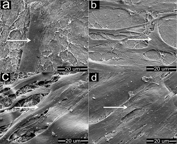
SHED is known for its capabilities to extensive proliferation and multipotential differentiation. The utilization of natural scaffold such as amniotic membrane, for SHED differentiation into odontoblast-like cells offers great potential in dental regeneration. This study aims to investigate the ultrastructure transformation during differentiation of SHED into odontoblast-like cells of SHED on AM in vitro using scanning electron microscope (SEM). SHED was cultured on AM scaffold and treated with bone morphogenetic protein-2 (BMP-2) growth factor for 1, 7, 10 and 14 days, respectively. SHED cultured on AM without BMP-2 served as control. Formaldehyde-based protocol was used to fix the samples for imaging. Samples were gold-coated with sputter coating machine SCD 0005 and viewed by SEM FEG450 at 5000x magnification. SEM imaging showed the surface structure of AM was fibrous. Nanofibrous structure is known to facilitate interaction of the cells for adherence and infiltration into this scaffold. SHED displayed cell extensions with small finger-like or web-like projections at the very end of the cell after a day of BMP-2 treatment. On the following days, SHED lost the fibroblast-like morphology, became smaller and round-shaped. Emergence of odontoblast-like cell with columnar cell body and several processes were detected on day 7. By day 14, strong mineralization on the cell body was observed, suggesting a matured odontoblast. In control group, SHED appearance as fibroblast-like cell morphology remained up until day 14 of culture. In conclusion, morphological evaluation via SEM confirms AM support for a complete differentiation of SHED into odontoblast-like cells by day 14 of culture.